内容摘要:
1,Resolution in STELLARIS
2,Why STED Super-Resolution?
3,Multicolor STED Imaging
4,Elucidating Protein Roles In Centriole Assembly
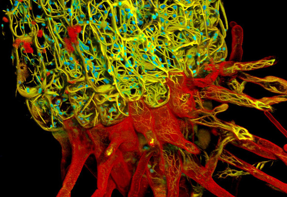
5,Monitoring Malaria Invasion Phase Using STED Imaging
6,Characterization of the glomerular barrier with 3D STED deep imaging
7,STED Imaging of Jurkat cells in 3D
8,Fluorescence imaging traditionally extracts spectral information
9,Fluorescence lifetime,荧光寿命
10,FLIM
11,Why is FLIM so powerfull
12,STimulated Emission Depletion is a Competing Process
13,What do we see with STED and lifetime imaging?
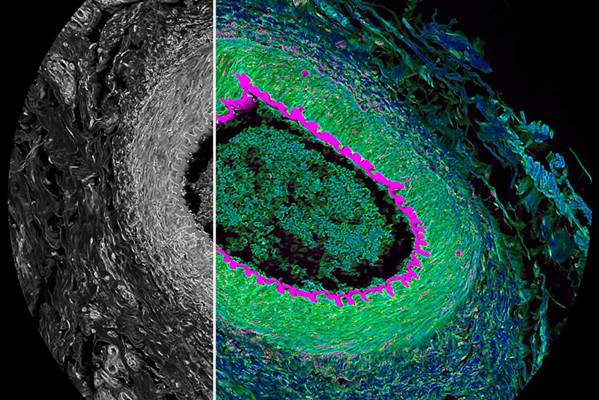
14,Tau STED delivers cutting-edge image quality and resolution
15,Tau STED imaging of Cytoskeleton components
16,Characterize transcriptional regulation with smFISH
17,3D STED on mammalian nuclear pore complex
18,3D STED on microtubule network
19,Tau STED 775 imaging of glomerular components in mouse kidney
20,STED with cutting edge optics: STED white objective lenses
21,Tau STED is gentle STED imaging for dynamic live cell experiments







* 该内容属于该讲师在职期间以徕卡显微系统公司员工身份,在徕卡显微系统公司出资举办/承办/赞助的线上/线下会议上做出的知识分享;
* 徕卡显微系统公司对该内容拥有著作权等相关权力。任何组织或个人,未经徕卡显微系统公司书面授权,严禁转载相关内容;
* 本页面中的讲师介绍基于该知识分享课程制作时的信息。随着讲师个人职业发展,相关讲师履历可能会有变化;
* 本课程中对相关技术、产品的描述内容仅指课程录制时的状态。随着技术的进步、产品的选代,相关描述可能不再适用于当下。如有疑问,可咨询徕卡显微系统官方客服热线。